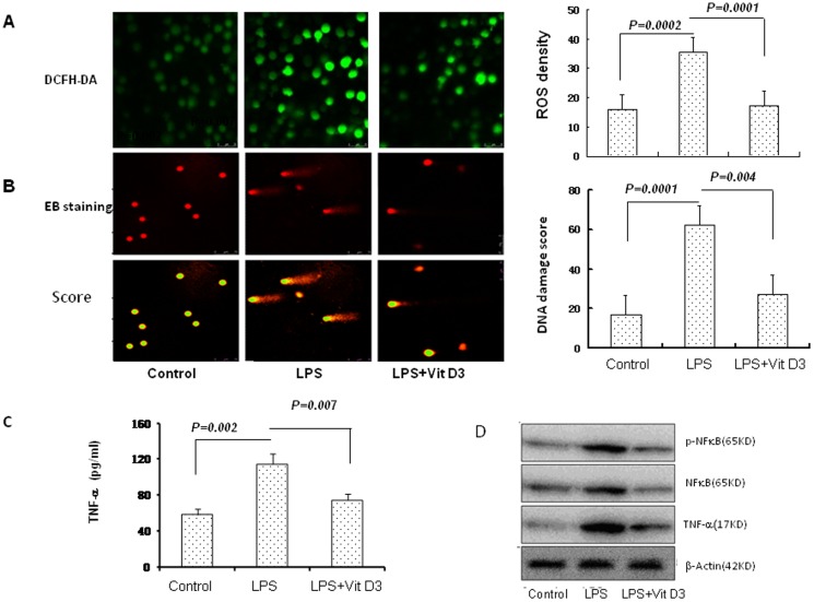
Figure 4. The effect of vitamin D3 on oxidative stress, DNA damage, TNF-α and NFκB in LPS-primed airway epithelial cells.
The ROS and DNA damage in airway epithelial cells was measured in the presence or absence of vitamin D3 and treated with LPS for 24 hours. A: ROS was detected by confocal microscopy using a DCFH-DA probe. The ROS level was represented by fluorescence intensity (X400). B: The DNA damage was measured by comet assay. The DNA damage score was analyzed with Comet Assay IV software. C: Standard ELISA was performed to determine the levels of TNF-α. D: NFκB were analyzed by western blot. β-actin was used as the loading controls.

