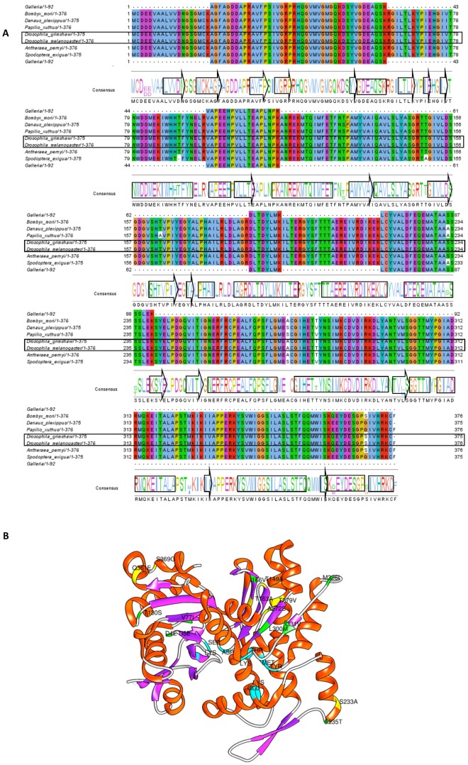
Figure 5. Actin 4 alignment and hypothetical 3D structure.
A. The actin alignment of Lepidoptera protein sequences matches that of G. mellonella's actin peptides (first discontinued sequences and dotted) and aligns well (100%) with the B. mori protein. Related species and Drosophila species were also included. A consensus was obtained on Jalview using a Clustal algorithm. The consensus sequence includes arrows (α helix) and cylinders (β-sheet) obtained by structural analysis. The alignment shows insertions, deletions or substitutions. B. Original tertiary 3MN6 (Protein Data Bank) actin pattern and probable amino acid substitutions are marked in yellow and green. The catalytic site residues can be found in blue.

