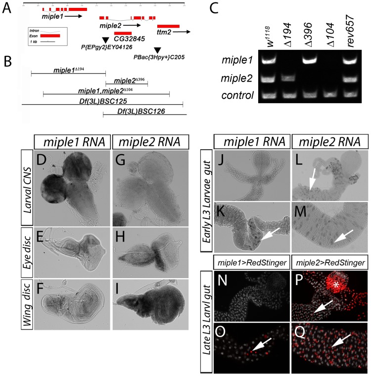
Figure 1. Generation of miple deletions and larval expression of miple1 and miple2.
(A) Schematic of the chromosomal region comprising the miple1 and miple2 loci on 3L with exons represented by boxes (translated regions depicted as broad red boxes; untranslated are dotted lines). The P-element employed for the imprecise excision screen, P{EPgy2}EY04126, is shown as filled inverted triangle. In addition to miple1 and miple2, two other genes – CG32845 (within the first intron of miple2) and ttm2 (downstream of miple2) are shown (red boxes). A putative ttm2 allele caused by the P-element PBac{3Hpy+}C205 upstream of ttm2 is shown as filled inverted triangle. (B) Regions deleted in the miple1Δ194 and miple2Δ396 single, and the miple1,miple2Δ104 double mutants are shown. Also indicated are the deletions in the deficiency lines Df(3L)BSC125 and Df(3L)BSC126. (C) Genomic PCR confirming gene deletion of miple1 and miple2 in the single and double mutants as indicated, controls are w1118 and revertant controlrev657. The control (lower band) was carried out to confirm genomic DNA quality. (D-I) In situ of miple1 and miple2 mRNA in larval tissues. Strong expression is detected in the larval optic lobe and CNS (D) and weak expression in imaginal eye (E) and wing discs (F). miple2 mRNA is not detectable in the larval CNS (G), while imaginal discs exhibit strong expression, represented by eye (H) and wing discs (I). (J-M) In early L3 larvae miple1 mRNA expression is detected in a distinct population of cells in the anterior midgut (K, see arrow) but not detected in other parts of the anterior midgut (J). Expression of miple2 mRNA expression is also detected in early L3 larvae in cells of the anterior midgut (L, see arrow) and posterior midgut (M, see arrow). The in situ expression pattern in the larval gut is mimicked by two Gal4 lines driving UAS-RedStinger (nuclear RFP, in red). miple1-Gal4 shows expression in a group of cells in the anterior part of the midgut (O, see arrow) but no expression in other parts of the gut (N), while miple2-Gal4 shows expression in AMPs in the larval midgut (P and Q, see arrow). Asterix (* in P) indicates ectopic Gal4 expression from observed in the proventriculus. Nuclear stain in (N-Q) visualises the gut (DAPI, white).

