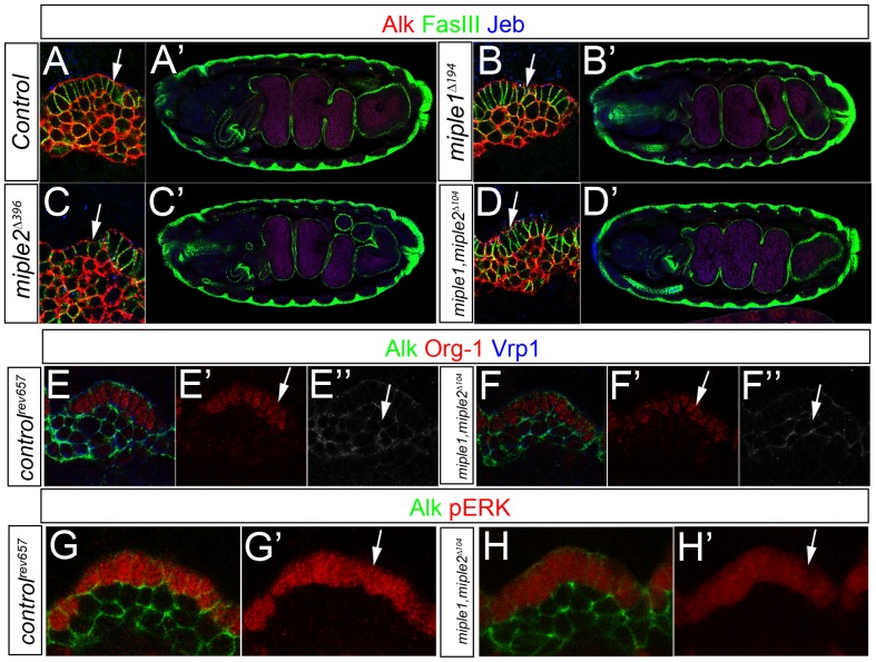
Figure 2. Miple proteins are not required for Alk signaling in the embryonic visceral mesoderm.
(A-D) The deletion mutants miple1Δ194, miple2Δ396 and miple1,miple2Δ104 exhibit normal visceral mesoderm development. In miple1Δ194, miple2Δ396 and miple1,miple2Δ104 deletion mutants, Alk positive columnar shaped founder cells (FCs, arrows) and fusion competent myoblasts (FCMs) are correctly specified (A, B, C, and D). At embryonic stage 17 midgut chambers are normally formed in all three mutants (B′, C′ and D′) and are comparable to control embryos (A′). Embryos are stained with Alk (in red), Jeb (in blue) and FasIII (in green). (E-F) At late stage 10 double miple1,miple2Δ104 deletion mutant embryos express FC (F′, Org-1 in red, arrow) and FCM (F′′, Vrp1 in white, arrow) specific markers as controlrev657 (E′, E′′). (G-H) At late stage 10, miple1,miple2Δ104 double mutant embryos display wild type ERK activity in FCs (H′, pERK in red, arrow) comparable with controlrev657 revertant control FCs (G, G′). Embryos are stained with Alk (in green) and Org-1 or pERK as indicated (in red).

