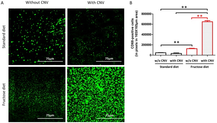
Figure 6. The number of CD68-positive cells is increased in the retina of rats fed with a 60%-rich fructose diet and submitted to laser-induced choroidal neovascularization (CNV).
A. Representative confocal images of CD68-positive cells (revealed by an Alexa 488-labelled secondary antibody) in flat-mounted retinas of rats fed during 3 months with either the standard or the 60%-rich fructose diet and submitted or not to laser-induced CNV. Images corresponding to 165 µm×165 µm of the retinal area were taken 3 weeks post laser-induced CNV. B. Quantification of CD68-positive cells in flat-mounted retinas of rats fed during 3 months with either the standard or fructose diet, and submitted or not to laser-induced CNV. ** statistically different at p≤0.01 (Kruskal-Wallis test). Values are expressed in pixels of fluorescence in the area corresponding to 165 µm×165 µm of the retina; values are given as means ± SD.

