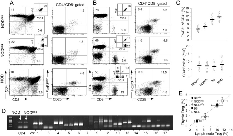
Figure 1. Efficient selection of CD4+ T lymphocytes in NODmini and NODβTg mice.
Lymphocytes isolated from thymii (A) and lymph nodes (B) of indicated mice were stained with monoclonal antibodies and analyzed by flow cytometry. Numbers in quadrants are representative percentages of at least six mice (6 week old) per group. The numbers of thymocytes and lymphocytes ± SD recovered from NODmini, NODβTg and NOD mice were: from thymii 78.5±16.7×10⁶, 72.05±13.8×10⁶ and 70.1±14.0×10⁶, and from lymph nodes (axillary, brachial and inguinal) 17.2±5.8×10⁶, 14.1±2.5×10⁶, and 13.2±3.0×10⁶, respectively. (C) Percentages (top) and total numbers (bottom) of CD4+Foxp3+ T cells in peripheral lymph nodes of 6 week old mice; each circle represents individual mouse. (D) Expression of mRNA of TCRVα genes in sorted CD4+ T cells isolated from NOD and NODβTg mice. Analysis was done by RT-PCR using primers specific to indicated Vα segments and Cα region. (E) Comparison of CD4+Foxp3+ T (Treg) cells from thymii and peripheral lymph nodes of transgenic and wild type B6 and NOD mice. Mean percentage and SD of six young mice per group are shown.

