Figure 6.
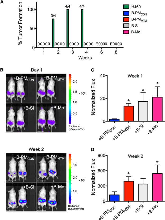
Chronic PMMTM exposed cells promote tumor formation of human nonsmall cell lung cancer H460 cells in mice. (A) Growth kinetics of H460 or transformed B-PMCON, B-PMMTM, B-Si, and B-Mo cells (1 × 10⁶ cells) when SC injected into the NSG mice alone. E indicates the end of experiment. (B) Transformed cells at the dose of 6 × 10⁵ cells were coinjected with luciferase-labeled H460 cells at the dose of 3 × 10⁵ cells (2:1 ratio) into the left and right flanks of NSG mice. Tumor formation was monitored weekly by IVIS bioluminescence imaging. Representative IVIS images of mice at day 1 and week 2 are shown. (C, D) Normalization of tumor bioluminescence signals at 1 (C) and 2 (D) weeks postinjection to their initial signal at day 1. Data are mean ± SD (n = 4). *P < 0.05 (power >60%) vs H460 and B-PMCON coinjection.

