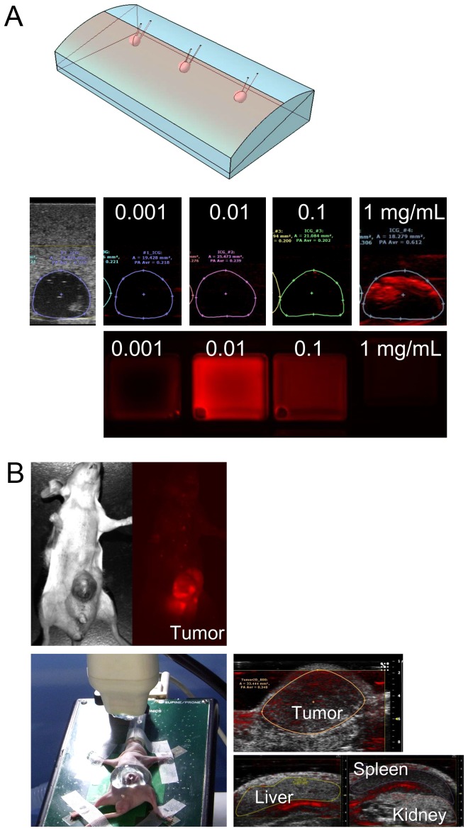
Figure 1. Establishment of PA tomography's ability to visualize ICG-containing tissue.
(A) Using a human liver tissue-mimicking phantom (top), human plasma containing ICG at concentrations of 0.001, 0.01, 0.1, and 1.0 mg/mL was encapsulated into holes that were 5 mm in diameter and located at a depth of 5 mm from the surface; PA amplitudes were measured using the Vevo LAZR imaging system (middle). Fluorescence images of each ICG-containing plasma sample were also obtained (bottom). (B) Fluorescence imaging in a mouse model with subcutaneously implanted well-differentiated human hepatoma cells (HuH-7) identified ICG accumulation in the subcutaneous tumor (left). PA tomography enabled differentiation of tumor-specific PA signals from those of surrounding organs under the conditions of 800-nm excitation light and 54-dB PA gain (right).

