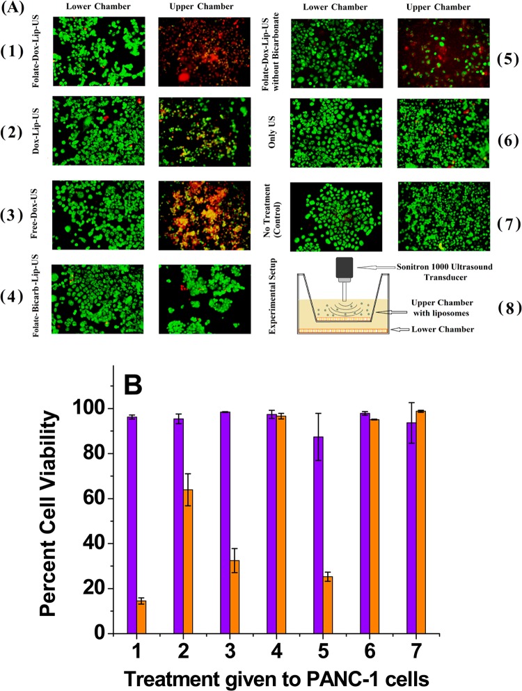
Figure 9.
(A) PANC-1 cell viability studies using live (green) and dead (red) cell staining of different treatment groups (n = 3). The upper chamber cells received direct exposure, whereas the lower chamber cells received indirect exposure to POPC liposomes and ultrasound. (1) Folate-targeted doxorubicin liposomes (encapsulating ammonium bicarbonate) + ultrasound. (2) Nontargeted doxorubicin liposomes (encapsulating ammonium bicarbonate) + ultrasound. (3) Free doxorubicin + ultrasound. (4) Folate-targeted liposomes (encapsulating ammonium bicarbonate but no doxorubicin) + ultrasound. (5) Folate-targeted doxorubicin liposomes (no ammonium bicarbonate encapsulation) + ultrasound. (6) Ultrasound only. (7) No treatment (control). (8) Schematic representation of the experimental setup. The final doxorubicin concentration used was 25 μg/mL. (B) Cell viability of the upper chamber (orange bars) and lower chamber (violet bars).

