
Figure 1.
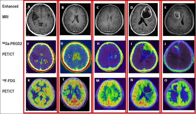
Demonstration of different WHO grade gliomas and comparison of the distribution of 68Ga-PRGD2 and 18F-FDG in the tumors. Enhanced MRI (upper row) of five patients was obtained after administration of gadolinium contrast agent. Low-grade gliomas (LGG) (A/F/K: F, 23 y, grade I. B/G/L: M, 40y, grade II) showed void to minimal accumulation of 68Ga-PRGD2 (middle row), whereas high-grade gliomas (HGG) (C/H/M: M, 41 y, grade III. D/I/N: M, 66 y, grade IV. E/G/O: M, 28 y, grade IV) showed moderate to intense uptake of 68Ga-PRGD2. The high-level cortical accumulation deteriorated the value of 18F-FDG PET/CT (lower row) in grading and demarcation of glioma, especially the LGG.