
Figure 2.
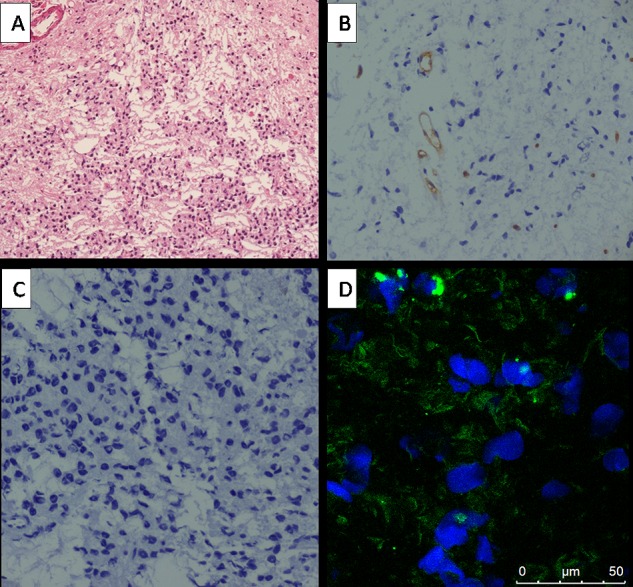
Immunohistochemical and immunofluorescence stains of the glioma of the patient (patient no. 1 Table 1, MRI and PET/CT images shown in Figure 1A,F,K) with LGG. (A) Hematoxylin-eosin staining showed WHO grade I glioma (magnification 100×). (B) The CD34 stains indicated few vascular networks in the tumors (magnification 200×). (C) Negative nuclear expression of Ki-67 indicated low proliferation (magnification 200×). (D) Very low expression of the integrin αvβ3 receptor were observed in the tumor or vascular endothelial cells.