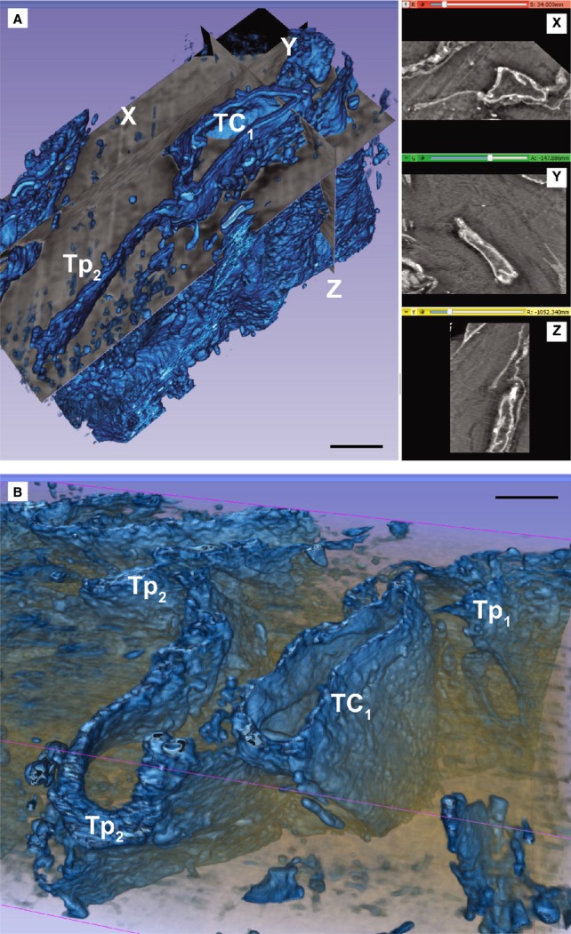
Figure 3.

(A and B) Automated segmentation of the stack containing the telocyte TC1 from Figure 2 shows that the telopode Tp2 is long (20 μm), narrow (0.2–1 μm) and flat, given a ribbon appearance of the cell. X-Y-Z slice projections from volume could be seen in the right side of A. Scale bars: 2 μm.
