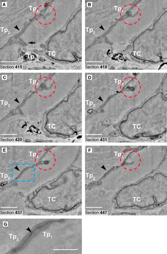
Figure 5.

(A–F) Six non-consecutive serial images (inverted) obtained in backscattered electron imaging mode at 150 nm z-interval. The quality of the images in FIB-SEM is comparable with classical transmission electron micrograph at 9kX magnification. The red ring indicates a characteristic dilation (podom) of the telopode Tp1, where intracellular structures such as endoplasmic reticulum cisternae and mitochondria are visible. A junction (processus adherens type) could be seen connecting the telopodes Tp1 and Tp2 (arrowheads). The area of this junction (rectangular marked area in E is enlarged in G) is about 5 μm2 (2 μm/2.5 μm). Another emerging junction (recessus adherens type) is visible (arrow in A) between telopodes Tp2 and Tp3 of the adjoining telocyte (TC). Scale bars: A–F, 2 μm; G, 1 μm.
