
Figure 5.
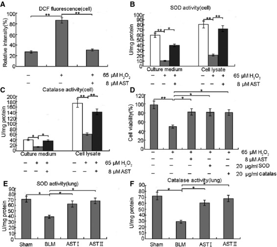
Antioxidative properties of AST facilitated protection from oxidative stress in vitro and in vivo. (A) RLE-6TN cells were treated with 65 μM H2O2 alone for 12 hrs and co-treated with 8 μM AST for 24 hrs. After treatment, ROS generation was determined using 5 μM CMH2DCFDA. AST exhibited significantly reduced H2O2-mediated ROS generation in RLE-6TN cells. (B and C) RLE-6TN cells were treated with 65 μM H2O2 alone for 12 hrs and co-treated with 8 μM AST for 24 hrs. SOD and catalase activities were measured after treatments. AST significantly increased SOD and catalase activities. (D) RLE-6TN cells were treated with 65 μM H2O2 alone for 12 hrs and co-treated with 8 μM AST, 20 μg/ml SOD or 20 μg/ml catalase for 24 hrs. AST, SOD and catalase improved cell viability. (E and F) The lung tissues were homogenated, centrifuged at 5000 × g for 15 min. at 4°C, and the supernatant was got to detect SOD and catalase. AST also increased SOD and catalase activities in vivo. *P < 0.05 and **P < 0.01, compared with the group treated with H2O2 or BLM alone.