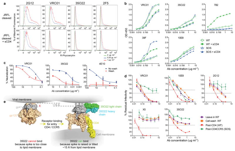
Figure 4. Binding or neutralization in the context of a lipid membrane.
a, Staining of cell surface expressed HIVJRFL Env. b, ELISA assay of antibody binding to WT or SOS HIVJRFL VLPs. The CD4-inducible 39F antibody or gp41-specific 7B2 are used as controls. c, Access to the HIVJRFL Env trimer on pseudovirions based upon washing the antibody-pseudovirion mixture prior to infecting cells. d, Kinetic assay of HIVJRFL neutralization. See Methods for a description of individual formats. e, Schematic of conformational change resulting in raising of the trimer spike required to permit access of 35O22 to its epitope.

