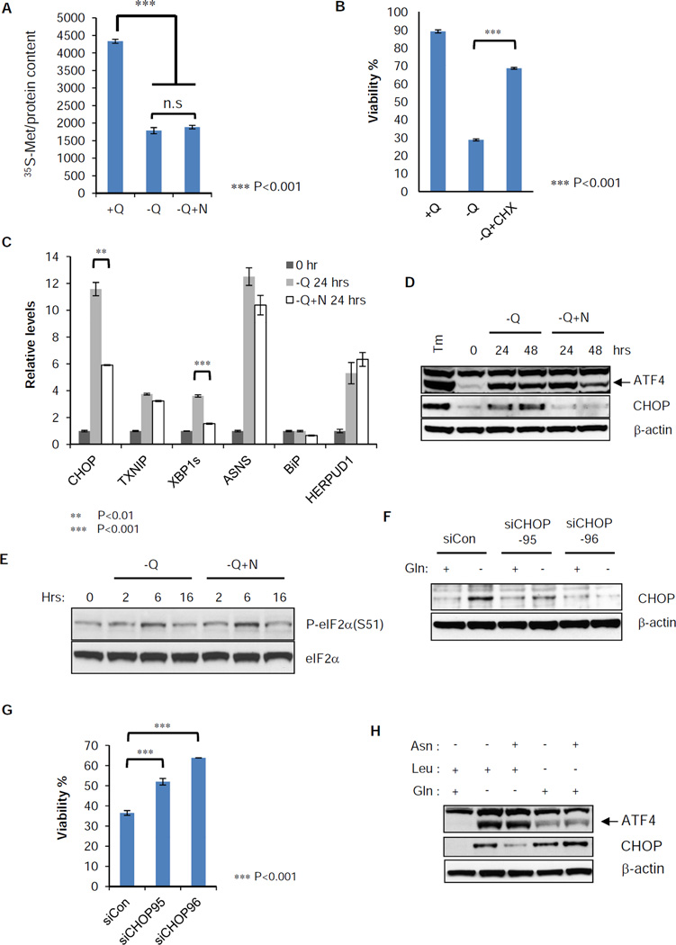
Figure 7. Induction of endoplasmic reticulum (ER) stress marker genes by glutamine withdrawal is suppressed by asparagine addition.
(A) SF188 cells were grown in complete medium (+Q) or glutamine-deficient medium with (−Q+N) or without (−Q) asparagine (4 mM) for 16 hours, then switched to the same fresh medium without methionine. 0.1 mCi 35S-methionine was pulsed for 30 minutes and whole cell extract was prepared. Radioactivity of labeled protein was measured in a scintillation counter and normalized to total protein content.
(B) SF188 cells were cultured 48 hours in DMEM with the following modifications: with glutamine (+Q), without glutamine (−Q) or without glutamine in the presence of cycloheximide (1 µg/mL) (−Q+CHX). Viability was measured by Annexin V and PI staining.
(C) SF188 cells were cultured in DMEM without glutamine (−Q) or without glutamine but with asparagine (4 mM) (−Q+N) for 24 hours. Q-VD was added at 20 µM to prevent cell death. mRNA was extracted and Q-PCR was performed to detect relative abundance of ER stress marker genes, CHOP, TNXIP, XBP1s, ASNS, BiP and HERPUD1 normalized to 18s ribosomal RNA.
(D) SF188 cells were cultured as in panel (C) for 48 hours. Protein extracts were prepared at 0, 24 and 48 hours for western blotting of ATF4 and CHOP. Tunicamycin (Tm) was added at 10 µg/ml for 4 hours as a positive control.
(E) SF188 cells were switched to minus glutamine DMEM with (−Q+N) or without (−Q) asparagine (4 mM) for 16 hours. Protein extracts were prepared at 0, 2, 6 and 16 hours after medium change. Western blotting was performed for phospho-eIF2α (S51) and total eIF2α.
(F) SF188 cells were transfected with control or CHOP siRNA for 2 days. Then glutamine was withdrawn for 24 hours in the presence of Q-VD (20 µM). Whole cell extracts were prepared for western blotting of CHOP.
(G) SF188 cells were transfected with control or CHOP siRNA for 2 days. Then glutamine was withdrawn for 48 hours, and viability was measured by Annexin V staining.
(H) SF188 cells were cultured in DMEM without glutamine (Gln) or leucine (Leu) individually. 4 mM asparagine (Asn) was added or not added to each amino acid deficient medium for 24 hours. Q-VD was added at 20 µM to prevent cell death and protein extract was prepared for western blotting of ATF4 and CHOP.
The data in Figure 7 (A, B, C and G) are shown as mean +/− S.D., n=3, and the p-values are determined by using Student’s 2 tailed t-test. See also Table S1.

