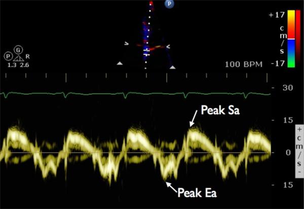
Figure 1.
Tissue Doppler velocities of the mitral annulus are shown for a sample patient. The sample volume is positioned over the septal annulus of the mitral valve. Peak systolic annular velocity (Sa) and early diastolic annular velocity (Ea) are indicated. Similarly, values for peak systolic myocardial velocity (Sm) and peak early diastolic myocardial velocity (Em) can be obtained by positioning the sample volume over a basal segment of the left ventricular wall

