
Figure 2.
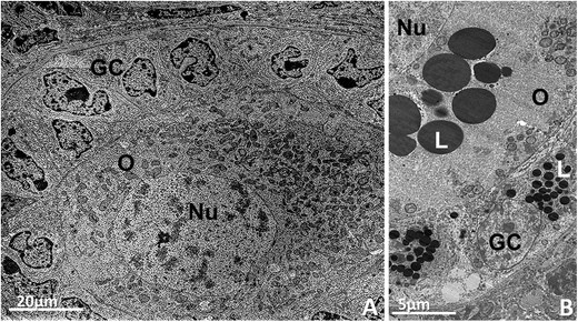
Primary follicles. A: Bovine primary follicle showing the oocyte with organelles homogeneally distributed throughout the cytoplasm surrounded by cuboidal granulosa cells. Round and elongated mitochondria can be observed. B: Pig primary follicle with several lipid droplets in the oocyte and granulosa cells cytoplasm. O: oocyte, Nu: nucleus, GC: granulosa cells, L: lipid droplet.