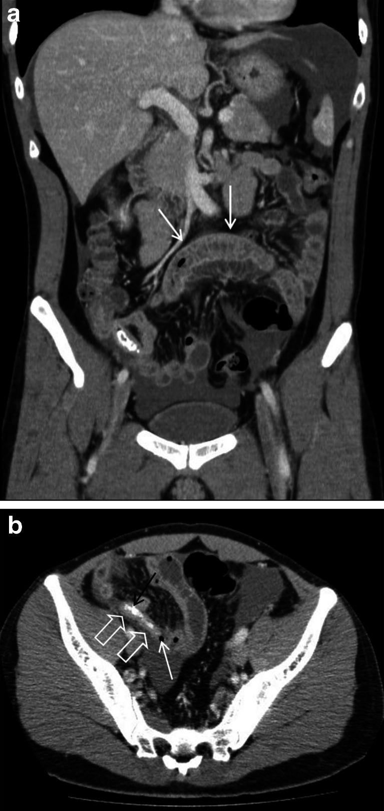
Fig. 2.
a A 32-year-old male with enteritis masquerading as acute appendicitis (case 2; see b). Contrast-enhanced coronal images demonstrate a fluid-filled small bowel loop with mural thickening and enhancement in keeping with enteritis (white arrows). Ascites is noted within the perisplenic region and pelvis. b A 32-year-old male with enteritis (case 2; see b). Contrast-enhanced axial CT image demonstrates a small air locule (white arrow) at the tip of a blind-ending tubular structure (open white arrows) within the right iliac fossa with a slightly thickened and enhancing wall, suggesting possible perforation of an acutely inflamed appendix. Intraluminal dense material was in keeping with an appendicolith (black arrow)

