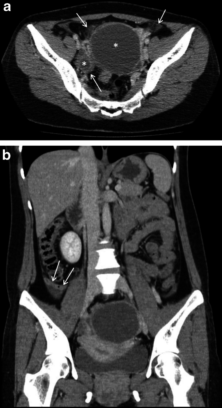
Fig. 3.
a A 35-year-old female with oophoritis in early pelvic inflammatory disease mimicking an acute appendicitis (case 3; see a). The contrast-enhanced axial CT scan image reveals incidental bilateral ovarian cysts, larger on the left (white asterisks). There are small pockets of free fluid and inflammatory fat stranding surrounding the ovarian cysts (white arrows). b A 35-year-old female with oophoritis in early pelvic inflammatory disease (case 3; see a). In this contrast-enhanced coronal CT image, there is a suggestion of mild focal mural thickening and enhancement of the retrocaecal appendix (white arrows), associated with a small amount of periappendiceal fluid

