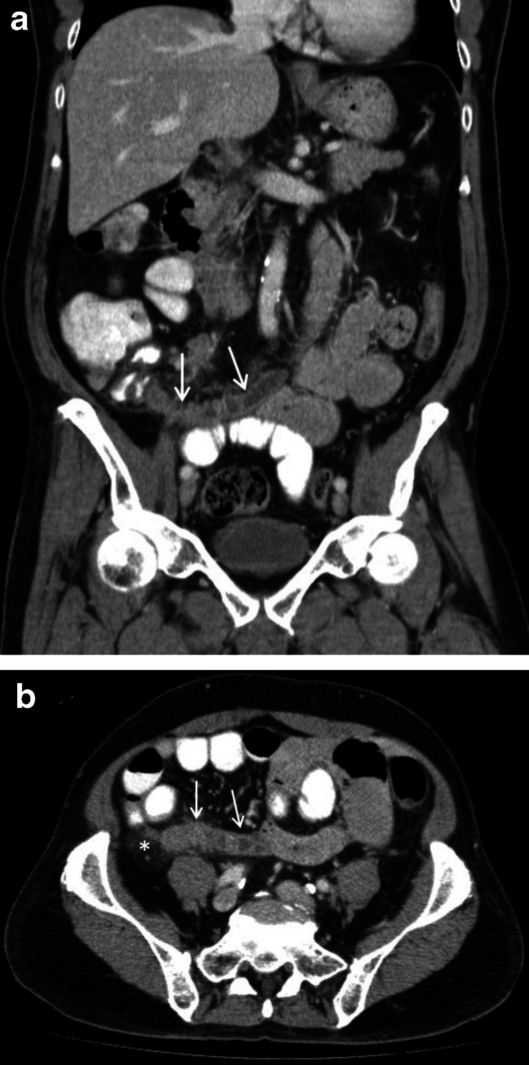
Fig. 4.
A 64-year-old male with moderately differentiated adenocarcinoma of the appendix (case 4). The coronal (a) and axial (b) contrast-enhanced CT scan images reveal a dilated, fluid-filled appendix with mural thickening and enhancement (white arrows). There is also evidence of periappendiceal inflammatory fat stranding (white asterisk) which is best appreciated in the axial image

