FIGURE 3.
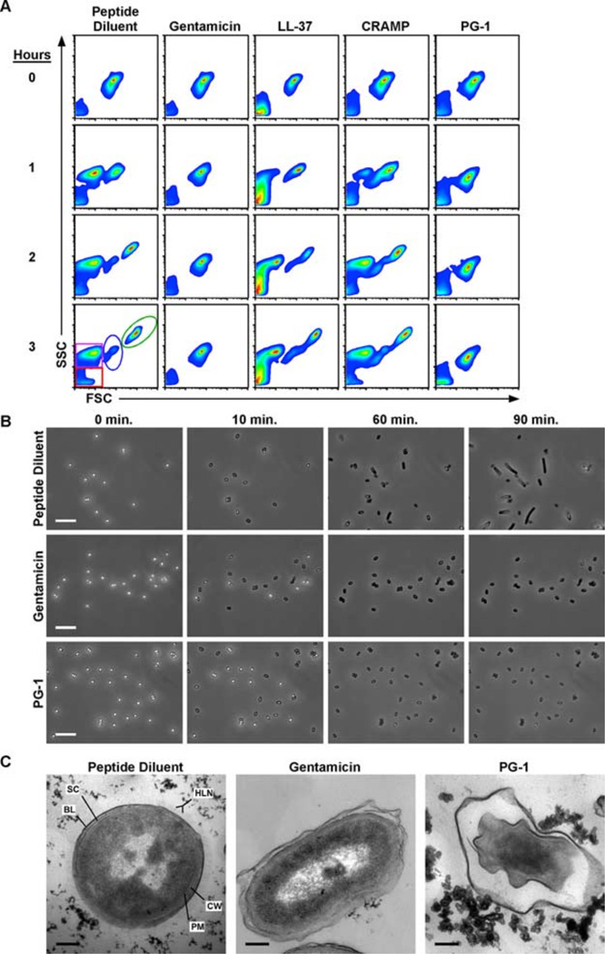
PG-1 treatment prevents vegetative outgrowth by disrupting the plasma membrane of the developing bacilli. In all experimental conditions, 1 × 10⁶ spores were incubated in peptide diluent control, genta-micin (5 µg/ml), or peptides (10 µM) diluted in MHB at 37°C for the indicated lengths of time. A, Flow cytom-etry was used to monitor the effects of each peptide on germination and outgrowth. Density plots depict the forward scatter (FSC-H) and side scatter (SSC-H) profiles of germinating spores in which debris (red), exosporium remnants (purple), spores (blue), and bacilli (green) can all be monitored as distinct populations. Scatter profiles are representative of three experiments conducted in duplicate. B, Representative photomicrograph excerpts taken from time lapse phase contrast microscopic analyses in which spores were incubated at 37°C and imaged every 10 s for 90 min. Photomicrographs are representative of three experiments. Bars = 10 µm. C, Representative transmission electron photomicrographs of spores treated with peptide diluent, gentamicin, or PG-1 for 1 h at 37°C. The plasma membrane (PM) and cell wall (CW) of the developing bacilli are shown in relation to the spore coat (SC) and basal layer (BL) and hair-like nap (HLN) of the exosporium. Bars = 0.2 µm.

