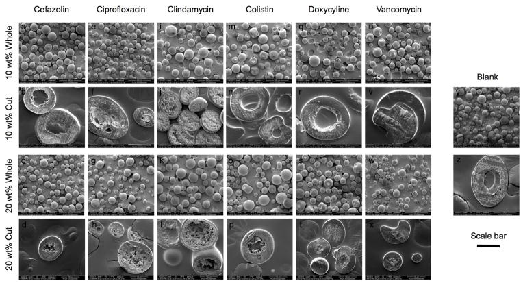
Figure 1.
Representative scanning electron micrographs of whole MPs and cut MPs to demonstrate external and internal morphology of cefazolin (a,b,c,d), ciprofloxacin (e,f,g,h), clindamycin (i,j,k,l), colistin (m,n,o,p), doxycycline (q,r,s,t), and vancomycin (u,v,w,x) of 10 wt% whole, 10 wt% cut, 20 wt% whole, and 20 wt% cut MPs, respectively. For comparison, blank MPs are shown whole and cut in (y) and (z), respectively. Whole MPs are shown at 100x magnification, and cut MPs are shown at 500x magnification. The scale bar represents 100 μm for rows 1 and 3 and (y), and 50 μm for rows 2 and 4 and (z).

