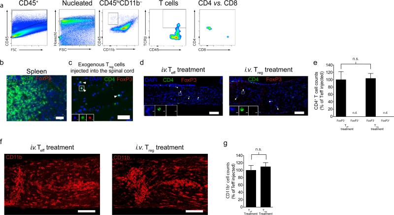
Figure 5. Boost with exogenous Treg cells does not alter immune cell infiltration into the injury site.
(a) Representative gates of flow cytometry of CD4+ and CD8+ lymphocytes in the injured optic nerve seven days post-injury. Optic nerves were pooled from eight mice, and cells were stained for analysis by flow cytometry. (b) Representative image from splenic tissue stained for CD4 (green) and Foxp3 (red) (scale bar = 100 μm). (c) Representative image from spinal cord tissue directly injected (ex vivo) with in vitro induced regulatory T cells stained for CD4 (green) and Foxp3 (red) (scale bar = 50 μm). (d) Representative images of CD4+Foxp3– and CD4+Foxp3+ T cells in the optic nerve parenchyma of Teff- and Treg-treated mice (scale bar = 100 μm). (e) Quantification of the number of CD4+Foxp3– and CD4+Foxp3+ T cells in the optic nerve parenchyma of Teff and Treg treated mice (n = 4 mice per group; One-way ANOVA with Bonferroni's post-test; scale bar = 100 μm). (f) Representative images of CD11b+ cells in injury site of the optic nerve of Teff- and Treg-treated mice (scale bar = 100 μm). (g) Quantification of the number of CD11b+ cells in injury site of the optic nerve of Teff and Treg treated mice (n = 9 mice per group; Student's t-test; representative of two experiments).

