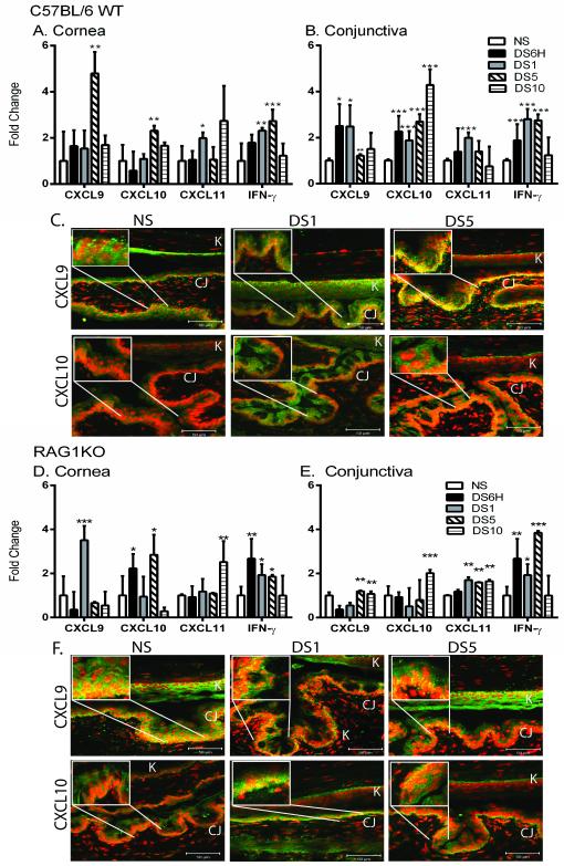
Figure 1. Induction of Th1-associated chemokines and IFN-γ occurs early in induction of dry eye disease.
A-B. Gene expression of CXCL9, CXCL10, CXCL11, and IFN-γ in cornea (A) and conjunctiva (B) of non-stressed (NS), six hours (DS6H) of desiccating stress (DS), one day of DS (DS1), five (DS5), or ten days (DS10) of DS C57BL/6 mice. C. Merged pictures of laser scanning immunofluorescent confocal microscopy of cornea and conjunctiva sections immunostained for CXCL9 (green) and CXCL10 (green) of NS, DS1, or DS5 of C57BL/6 WT mice with propidium iodide (PI) (red) nuclear counter staining. Yellow indicates strong double positive staining. D-E. Gene expression of CXCL9, CXCL10, CXCL11, and IFN-γ in cornea (E) and conjunctiva (F) of NS, DS6H, DS1, DS5, or DS10 of RAG1KO mice. F. Merged pictures of laser scanning immunofluorescent confocal microscopy of cornea and conjunctiva immunostained for CXCL9 (green) and CXCL10 (green) of NS, DS1, or DS5 of RAG1KO mice with propidium iodide (red) nuclear counter staining. Scale bar −50 μm. Results represent the mean ± SD expression of 6-8 animals per time point.*, p < 0.05; **, p < 0.01; ***, p <0.001 compared to NS. White boxes indicate magnified area.

