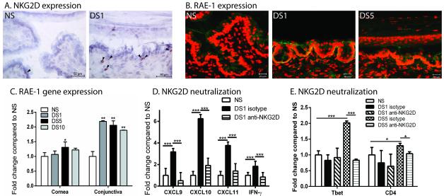
Figure 5. NKG2D and its ligand, RAE-1, increases with exposure to DS and NKG2D neutralization decreases expression of Th1-associated chemokines and IFN-γ.
A. Immunohistochemistry staining of NKG2D (red) with black arrows in the conjunctiva of NS and DS1 C57BL/6 mice B. Merged pictures of laser scanning immunofluorescent confocal microscopy of cornea and conjunctiva sections immunostained for RAE-1 (green) in conjunctiva of NS, DS1, or DS5 WT C57BL/6 mice. Counter stained with PI (red). Scale bar −20 μm. C. Gene expression of RAE-1 in cornea and conjunctiva of non-stressed (NS), one day (DS1) of desiccating stress (DS), five days of DS (DS5), or ten days (DS10) of DS WT C57BL/6 mice. D. Neutralization of NKG2D decreases conjunctival expression of CXCL9, CXCL10, CXCL11, and IFN-γ. E. Gene expression of Tbet and CD4 in the conjunctiva of non-stressed (NS), one day (DS1) of desiccating stress (DS), five days of DS (DS5), or ten days (DS10) of DS WT C57BL/6 mice. Results represent the mean ± SD expression of 6-8 animals per time point.*, p < 0.05; **, p < 0.01 compared to NS.

