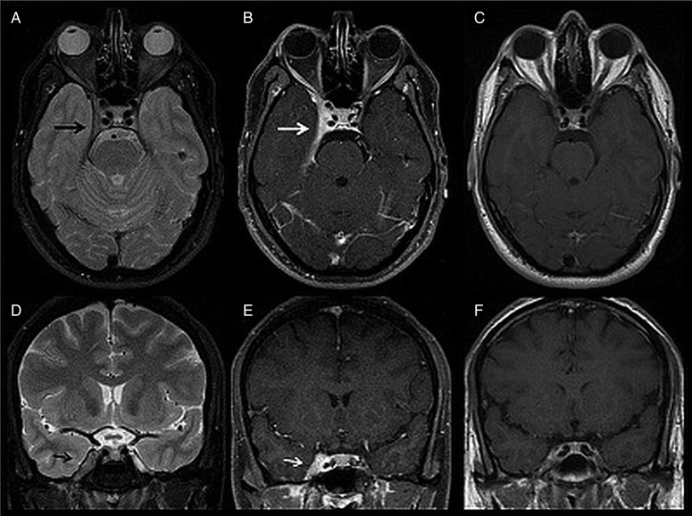
Figure 1.
MRI finding of a 36-year-old male patient with right-sided Tolosa-Hunt syndrome. Axial (A) and coronal (D) T2-weighted images (WI) show an enlarged right cavernous sinus (CS) that is mildly hypointense to grey matter (black arrow). Axial (B) and coronal (E) postcontrast T1-weighted fat-suppressed images show the enhancement of the abnormal soft tissue extending through the superior orbital fissure into the orbital apex (white arrow). Also noted is the hyperenhanced thickening of the right temporal dura, tentorium and right orbital apex. Axial (C) and coronal (F) postcontrast T1-WI follow-up, performed one month later, show significant improvement of right-sided CS abnormal enlargement and enhancement.

