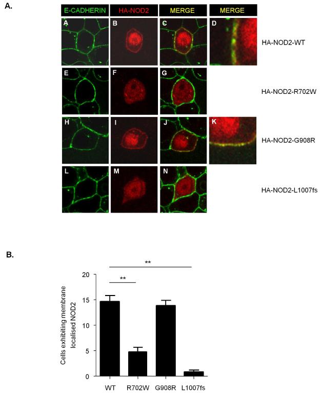
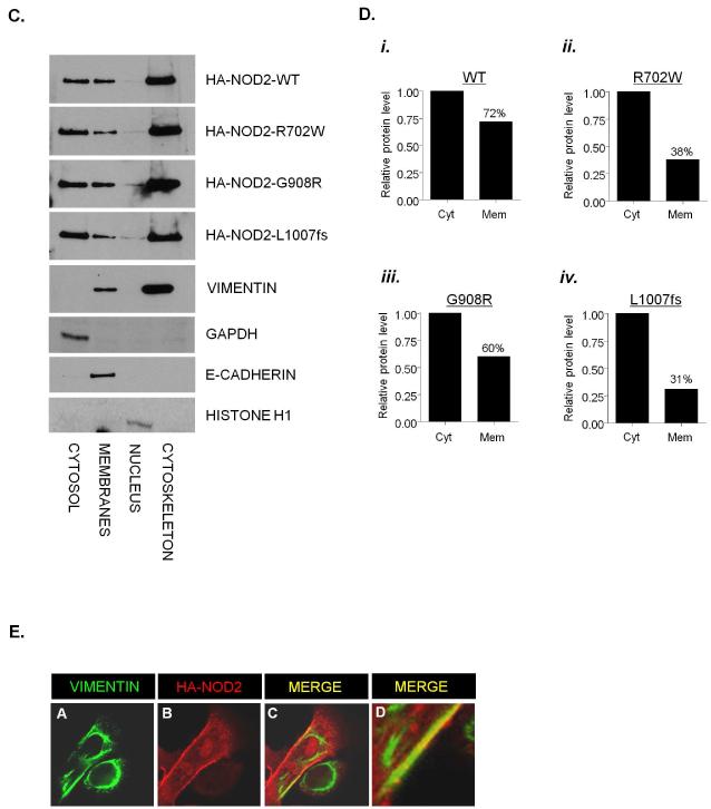
Figure 3. Subcellular localisation of CD-associated NOD2 variants.
(a) HEK293 cells were transfected with HA-NOD2, HA-NOD2-R702W, HA-NOD2 - G908R or HA-NOD2-L1007fs variants and immunostained with E-cadherin specific antibodies (green) and exogenous NOD2 with HA-11 antibodies (red). Merged images demonstrate areas where NOD2 and E-cadherin co-localise (yellow) and are highlighted in HA-NOD2 and HA-NOD2-G908R cells (panels d,k).
(b) Quantification of NOD2 membrane localisation in Figure 2A. Twenty cells from three separate fields of view were evaluated for membrane localized NOD2. *** = P<0.001.
(c) HEK293 cells were transfected with HA-NOD2, HA-NOD2-R702W, HA-NOD2 - G908R or HA-NOD2-L1007fs variants. Following transfection, proteins were separated into specific subcellular fractionations. Each fraction was immunoblotted for NOD2 proteins, vimentin, GAPDH, E-cadherin and Histone H1.
(d) Relative protein membrane fraction of HA-NOD2 (i), HA-NOD2-R702W (ii), HA-NOD2-G908R (iii) and HA-NOD2-L1007fs (iv) in relation to the cytosolic fraction using densitometry. Cyt, cytosol; Mem, membrane.
(e) HEK293 cells were transfected with HA-NOD2 and immunostained with HA-11 antibodies (red) vimentin-specific antibodies (green). Areas where NOD2 and vimentin co-localise are highlighted and appear as yellow.


