
Fig 6.
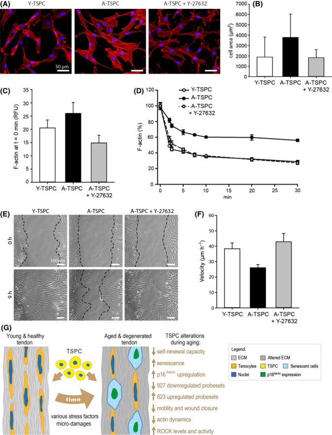
ROCK inhibition in A-TSPC. (A) Phalloidin staining of Y-TSPC, A-TSPC, and A-TSPC treated with the ROCK inhibitor 10 μm Y-27632. (B) Quantification of cell area. TSPC were cultivated on collagen I (200 cells per donor group were analyzed). (C) Comparison of F-actin content of Y-TSPC, A-TSPC, and A-TSPC + Y-27632 prior LanA treatment. RFU, relative fluorescent units. (D) Quantification of the F-actin amount upon LatA treatment. (E and F) Scratch assays on collagen I. Representative images at 0 and 9 h; cellular fronts are outlined with black lines. Cell velocity was calculated from four scratches per donor. The above-mentioned experiments were reproduced twice independently with three different donors per group. Bar charts show mean ± SD. (G) Schematic summary of the major findings detected in TSPC during tendon aging and degeneration. Young and healthy tendon tissue consists of abundant extracellular matrix, terminally differentiated tenocytes and resident stem and progenitor cells (TSPC). During aging, various stress factors and mechanical microdamages lead to altered tendon composition and functionality. Here, we report that in this process, TSPC accumulate several profound changes which altogether result in the exhaustion of their number and functional fitness.