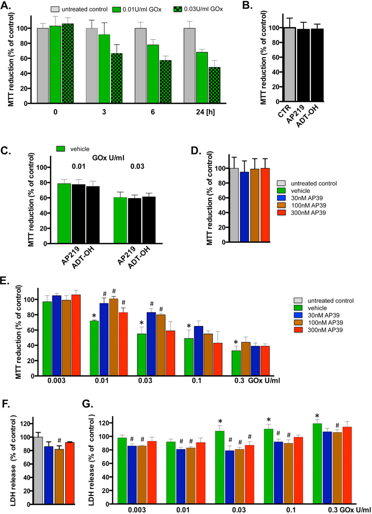
Fig. 5. Cytoprotective effects of AP39 in oxidatively stressed cells.
(A) Time-course of the change in MTT reduction at various time points after glucose oxidase exposure in bEnd.3 endothelial cells. Cells were exposed to glucose oxidase (0.01 or 0.03 U/ml) for 1h, followed by a washout and replacement of the medium with fresh tissue culture medium. Cells were incubated for a subsequent 23h period, followed by the measurement of MTT conversion. (B) Lack of effect of AP219 or ADT-OH (100 nM) on MTT reduction in bEnd.3 endothelial cells, as measured at 24 hours. (C) Lack of protective effect of AP219 or ADT-OH (100 nM) on the glucose oxidase-induced decrease in MTT reduction, as measured in bEnd.3 endothelial cells at 24 hours. (D) Lack of effect of AP39 (30–300 nM) on the mitochondrial conversion of MTT to formazan, an index of mitochondrial function/cell viability in bEnd.3 endothelial cells at 24 hours. Note that AP39 alone does not affect MTT conversion. (E) Effect of various concentrations of AP39 on MTT conversion in cells exposed to various concentration of oxidative stress induced by increasing concentration of glucose oxidase (GOx). Note that there is a decrease in MTT conversion in GOx treated cells; these effects are attenuated by AP39. Please also note that AP39-mediated protection was only observed at intermediate concentrations of GOx; the protective effects were no longer observed at the highest concentrations (0.1 – 0.3 U/ml) of GOx used. (F) Effect of AP39 on the breakdown of the integrity of the plasma membrane, as measured by LDH release into the extracellular medium. Please note that an intermediate concentration of AP39 attenuates basal LDH release in cells not treated with oxidants, perhaps indicative of improved viability or protection against a small degree of baseline cell dysfunction/cell death. (G) Effect of AP39 on LDH release in cells exposed to various concentration of GOx. Please note that AP39 decreased the release of LDH at intermediate concentrations of GOx; the protective effects were no longer observed at the highest concentration (0.3 U/ml) of GOx used. * p<0.05 shows a significant decrease in MTT or a significant increase in LDH in response to GOx treatment, when compared to baseline control (in the absence of GOx or AP39). # p<0.05 shows a significant enhancement of MTT or a significant reduction of LDH by AP39, when compared to its corresponding control at the same concentration of GOx, or in the absence of GOx. Results shown in parts A–G show mean±SEM values from three independent experiments with 4–8 replicates for each end-point.

