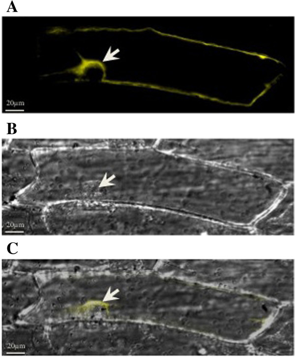
Figure 5.

Subcellular localization of TaSUT2 transiently expressed in onion epidermal cells. Localization of the TaSUT2-YFP fusion protein to the tonoplast is shown by the white arrow (A). Differential interference contrast (DIC) image of the same onion epidermal cell (B); the white arrow indicates the nucleus of the cell. Merged images of A and B to co-localize the TaSUT2-YFP to the tonoplast (C). No YFP-fluorescing signal was detected in the negative control (data not shown).
