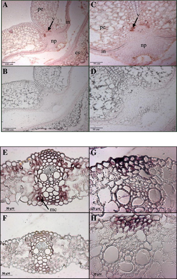
Figure 9.

Cellular localization of TaSUT2 transcripts in wheat seeds and leaves. Transverse sections of the middle portion of 4 DAA wheat seeds probed with digoxygenin-labeled antisense (A, C) and sense (B, D)TaSUT2 riboprobes. The TaSUT2 transcripts are mainly localized to the vein (see the red-brown staining indicated by the arrow in A, C). Weak signal (light pink staining) was also detected in the tip of the nucellar projection and in the integument (A, C). A and B, and C and D are at 5X and 10X magnifications, respectively. Transverse sections of the youngest fully expanded leaf of 1-month-old wheat plant probed with digoxygenin-labeled antisense (E, G) and sense (F, H)TaSUT2 riboprobes. The TaSUT2 transcripts are mainly localized to the subepidermal mesophyll cells (see the red-brown staining indicated by the arrow in E). Weak signal (light pink staining) was also detected in the vein (E, G). E and F, and G and H are at 10X and 20X magnifications, respectively. Transverse sections hybridized with the control sense probe (B, D and F, H) produced no signal. v, vein; np, nucellar projection; in, integument; es, endosperm; pe, pericarp; mc, mesophyll cells.
