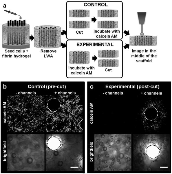
Figure 3.
Diffusion of calcein AM into the scaffold bulk. (a) Unchanneled and channeled scaffolds (height=6 mm, ø=12 mm) were seeded with human dermal fibroblasts and cultured statically. One group of scaffolds was cut down the centerline and stained with calcein AM (control, pre-cut). Another group was not sliced down the centerline prior to staining with calcein AM and therefore any positive staining in the experimental condition resulted from diffusion of the calcein AM molecule into the bulk of the scaffold (experimental, post-cut). (b) Representative control samples showing presence of live cells in the middle of unchanneled and channeled scaffold. (c) Representative experimental samples showing calcein AM diffusion into the middle of channeled, but not unchanneled scaffolds. Average radius of calcein AM diffusion around the channels as determined by Image J analysis was 321.7±13.5 μm. Scale bars=300 μm.

