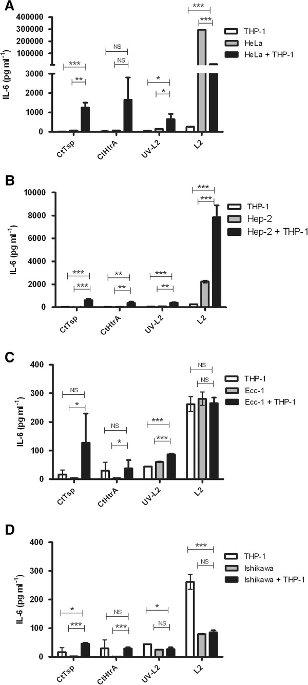
Figure 1.

IL-6 production by laboratory model epithelial cells and THP-1 cells to chlamydial antigens and Chlamydia. The graphs show the supernatant concentration of IL-6 detected at 96 hrs after cells were stimulated with CtTsp, CtHtrA, UV killed Chlamydia, or live Chlamydia (as labelled on the x-axis). HeLa cells A) are of cervical origin, HEp-2 cells are derived from male epidermoid cells B), while Ecc-1 C) and Ishikawa D) cells are of endometrial origin. Each cell model was also co-cultured with THP-1 (culture conditions indicated by the legend to top left of each graph). The IL-6 values were corrected for mean baseline IL-6 levels seen in unstimulated cells. Unpaired two-tailed t-tests have been performed (n = 3). NS = not significant. * p < 0.05, **p < 0.01, *** p < 0.001.
