
Figure 16.
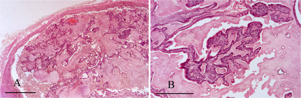
Renal papillary adenoma. A: Low magnification image of kidney showing proliferating cords of epithelial cells within a cystic structure that is compressing the adjacent parenchyma. The tissue is markedly autolysed. H & E Stain, bar = 1 mm. B: Higher magnification image of the same kidney section showing a proliferating, frond-like cord of the tumour. H & E Stain, bar =200 mμ.