
Figure 6.
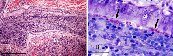
Bordetella bronchopneumonia. A: Histological section of lung showing a bronchiole occluded by a mass of inflammatory cells within a mucofibrinous matrix. H & E Stain, bar = 300 mμ. B: High power view of lung showing masses of B. bronchiseptica organisms (arrows) adhering to bronchiolar epithelium. Giemsa stain, bar = 25 mμ.