
Figure 4.
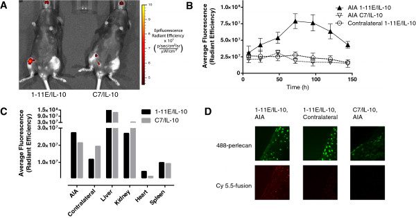
Tracking of 1-11E/vIL-10 to arthritic joints. One day after AIA induction in mice, 1 μg of Cy5.5-labeled fusion proteins was injected i.p. In vivo fluorescence images of the mice were taken to follow the tracking of the fusion proteins to the joint. (A) Representative fluorescence image taken 3 days after injection of 1-11E/vIL-10 (left) and C7/vIL-10 (right). (B) Quantification of the average fluorescence of the region of interest encompassing the knee joint, n = 3. (C)Ex vivo quantification of fluoresce of tissues after dissection of a single mouse 4 days after injection. (D) Representative fluorescence images of cryosections of the excised knee joints, showing the pericellular matrix of the chondrocytes in green (AlexaFluor-488-labeled secondary) and the fusion protein (Cy5.5) in red.