
Fig. 7.
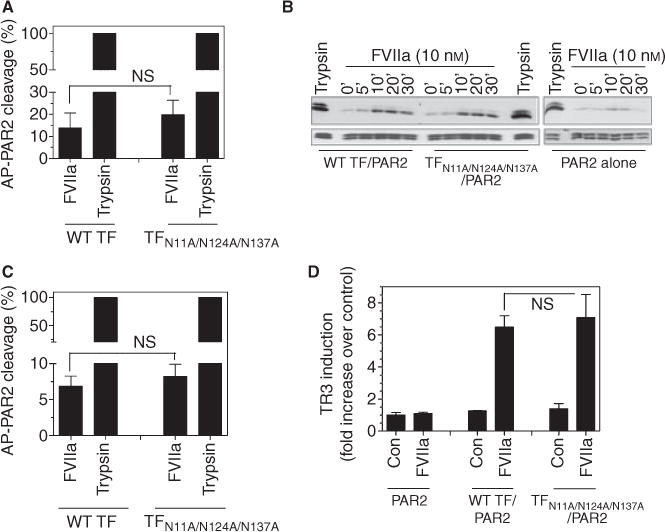
Factor (F) VIIa cleavage of PAR2 and activation of p44/42 MAPK in cells expressing wild-type or non-glycosylated tissue factor (TF). (A and B) CHO cells were co-transduced with 100 MOI per cell of wild-type TF-GFP and TFN11A/N124A/N137A-GFP adenovirus with 25 MOI per cell of AP-PAR2 adenovirus or with AP-PAR2 alone for 48 h. (A) The transduced cells were incubated at 37°C with a control vehicle, FVIIa (10 nM) or trypsin (5 nM), and at the end of 1 h incubation, soluble alkaline phosphatase activity released in the medium was measured. The values obtained in control vehicle treatment were subtracted from FVIIa and trypsin treatments and the value obtained in trypsin treatment was designated 100% of PAR2 cleavage. (B) Cells deprived of serum overnight were treated with FVIIa (10 nM) for varying times (0–30 min) or trypsin (5 nM) for 5 min and the cell lysates were subjected to SDS-PAGE and immunoblotted with phospho p44/42 or total MAPK antibodies. (C) Human umbilical vein endothelial cells (HUVEC) were infected with 20 MOI per cell of wild-type TF-GFP or TFN11A/N124A/N137A-GFP adenovirus and 10 MOI per cell of AP-PAR2 adenovirus. Cells were treated with FVIIa and trypsin and cleavage was determined as described for panel A. (D) HUVEC were transduced with PAR2 (10 MOI per cell) and either wild-type TF-GFP or TFN11A/N124A/N137A-GFP (20 MOI per cell) adenovirus for 48 h. The cells were serum starved for 2 h in EBM-2 SFM and then treated with FVIIa (10 nM) for 90 min. Total RNA was isolated and subjected to real-time qPCR analysis in triplicates. Fold-increase in TR3 mRNA levels was measured relative to TR3 mRNA levels in cells expressing PAR2 (no TF) and treated with control vehicle. ns, no statistically significant difference.