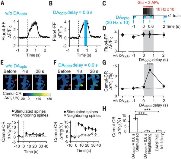
Fig. 3. DAopto effects on STDP stimulation-induced increases in [Ca2+]i and CaMKII activities.
(A and B) Increases in Fluo4-FF fluorescence, representing [Ca2+]i increases, within single spines in response to a train of STDP stimulation in the absence [(A), 20 spines, 15 dendrites] and presence of DAopto with a 0.6-s delay [(B), 15 spines, 8 dendrites]. Blue laser irradiation during DAopto is blanked by the blue bar. (C) STDP and DAopto protocols for Ca2+ and CaMKII imaging. Unlike plasticity induction (Fig. 1N), only one train was applied. (D) No effect of DAopto on the peak values of [Ca2+]i (fig. S6, A to C). P = 0.91 with Kruskal-Wallis test. (E and F) Ratiometric imaging with Camuiα-CR during STDP stimulation in the absence [(E), 33 spines, 14 dendrites] or presence of DAopto with a 0.6-s delay [(F), 42 spines, 14 dendrites]. Relative increases in the ratio are shown as pseudocolor coding in (E). (Bottom) Time courses of FRET ratios in the spines stimulated with glutamate uncaging or the neighboring spines. Scale bars, 1 μm. (G) Dependence of the peak Camuiα-CR FRET ratios on the DAopto delay (fig. S6, D to F). P = 1.3 × 10⁻⁵ with Kruskal-Wallis test and ***P = 8.4 × 10⁻⁵ with Steel test versus those without DAopto. (H) Normalized increases in Camuiα-CR ratios by STDP + DAopto with a 0.6-s delay in the stimulated (42 spines, 14 dendrites) and neighboring spines (42 spines, 14 dendrites), and in the presence of DARPP-32 inhibitory peptide in the stimulated spines (43 spines, 10 dendrites) (fig. S6G). Data are presented as mean ± SEM. P = 8.8 × 10⁻⁹ with Kruskal-Wallis test and *** P = 2.6 × 10⁻⁹ and 1.1 × 10⁻⁶ with Steel test.

