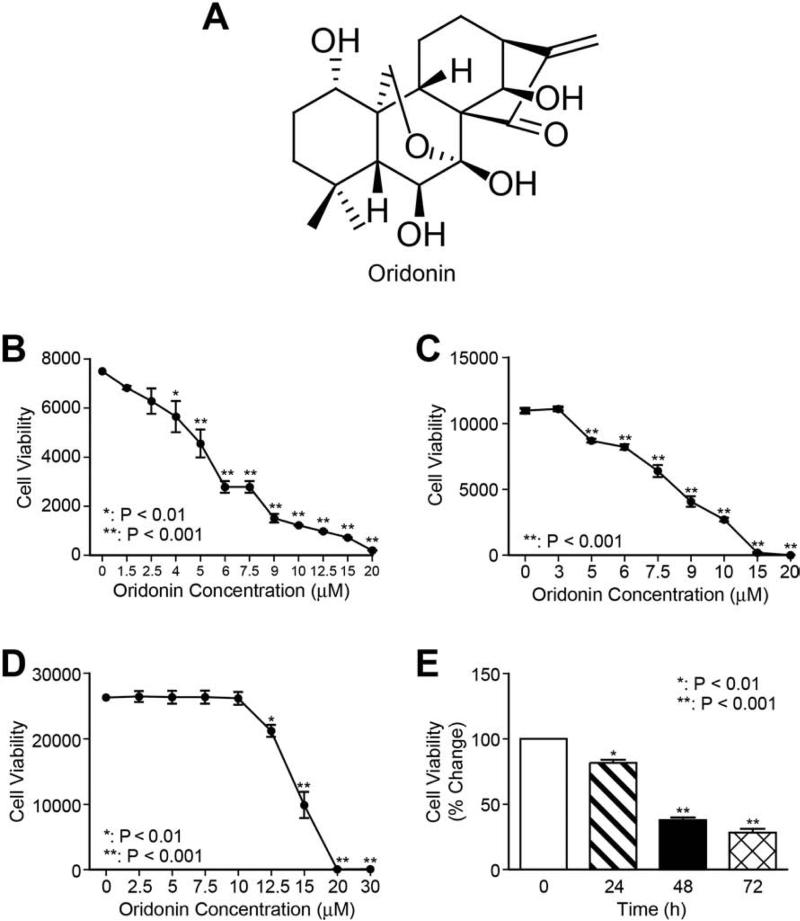
Fig. 1. Oridonin suppresses HSC proliferation.
Chemical structure of oridonin (A). LX-2 cells (B), HSC-T6 cells (C) and C3A hepatocytes (D) were treated with a series of concentrations of oridonin for 48 hours, and cell viability was determined using Alamar Blue assay. (E) LX-2 cells were treated with 7.5 μmol/L of oridonin for 24, 48, and 72 hours; cell viability was measured by Alamar Blue assay. P-values shown compared to vehicle (0.1% DMSO, 0 μmol/L). The results are representative of at least three independent experiments.

