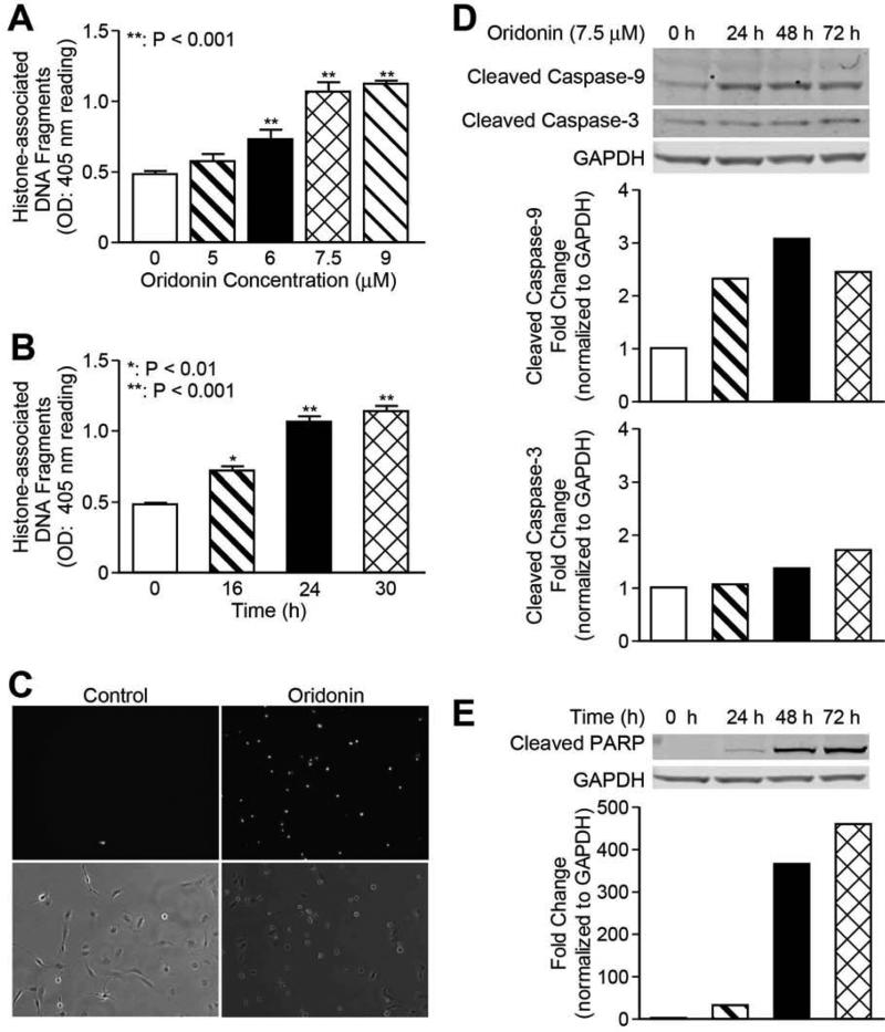
Fig. 3. Oridonin promotes LX-2 cell apoptosis.
LX-2 cells were incubated with different concentrations or time points as indicated. Apoptosis by oridonin was evaluated either by Cell Death detection ELISA (each conducted in triplicate) (A) and (B), or Yo-Pro-1 staining (C). Whole cell lysates were analyzed by Western blot with antibodies for cleaved caspase-3 and cleaved caspase-9 (D), cleaved-PARP (E). GAPDH was used as loading control. Densitometric analyses of bands were quantified and data expressed as fold of control normalized to GAPDH. The results are representative of at least three independent experiments.

