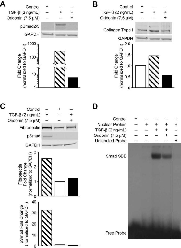
Fig. 5. Oridonin inhibits TGF-β induced pSmad activity and ECM.
LX-2 cells were preincubated with oridonin (7.5 μmol/L) for 2 hours and then treated with TGF-β (2 ng/mL) for 18 hours, whole cell lysates were analyzed by Western blot with antibodies for phosphorylated-Smad2/3 (A) and type I collagen (B), fibronectin (C). GAPDH was used as loading control. Densitometric analyses of bands were quantified and data expressed as fold change of control normalized to GAPDH. (D) LX-2 cells were pretreated with 7.5 mol/L of oridonin for 1 hour and then treated with TGF-β (2 ng/mL) for 1 hour. Nuclear proteins were extracted and analyzed by EMSA with 32P-labeled SBE probe described in experiment procedures.

