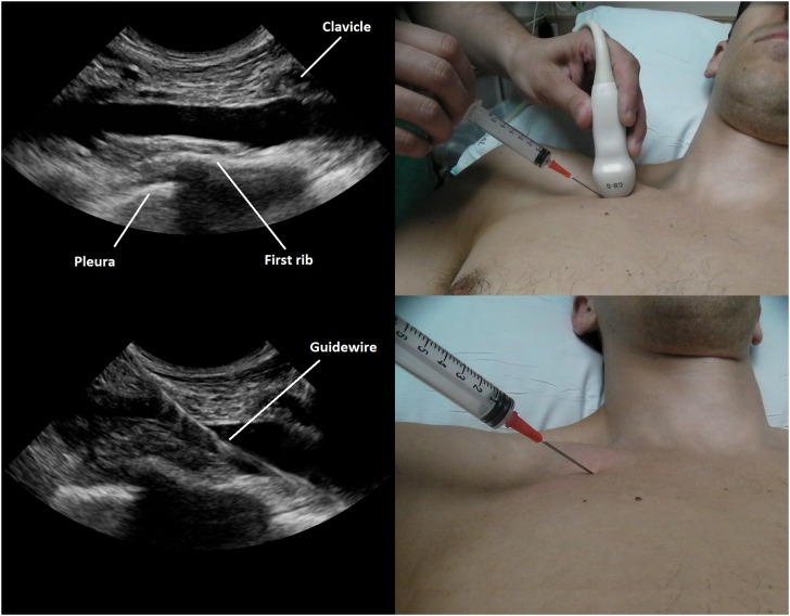
Figure 2.
Ultrasound image of a right subclavian cannulation. The probe is placed at the inferior margin of the clavicle (top right), and the subclavian vein is imaged (top left). The needle is placed directly under the curved portion of the probe, entering at a 45°angle (top right). The guidewire is seen entering the subclavian vein. Subclavian placement is confirmed because the needle and wire entry are medial to the lateral margin of the first rib (bottom left). This method allows for needle entry within 1 to 2 cm from the clavicle (bottom right).

