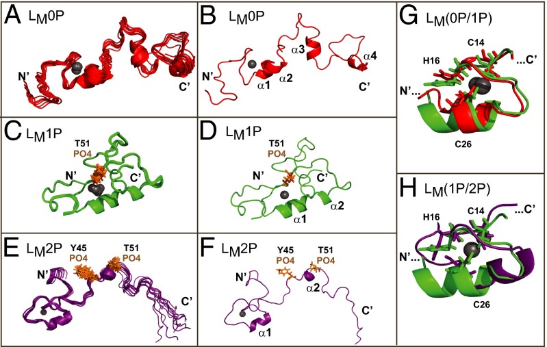
Fig. 2.
Solution structures of LM(0P/1P/2P). (A, C, and E) The 10 lowest-energy states for LM0P, LM1P, and LM2P as free solution structures are shown. These ensembles are as deposited with PDB. (B, D, and F) The state-1, lowest energy structure for each protein is labeled with determined motifs. (G) Superimposition of the zinc-finger regions of LM0P and LM1P highlight observed rearrangements. (H) Similarly, superimposition of LM1P and LM2P zinc finger regions show conformational changes centering on the zinc binding domain, particularly H16. In all panels, the zinc ion is a gray sphere.

