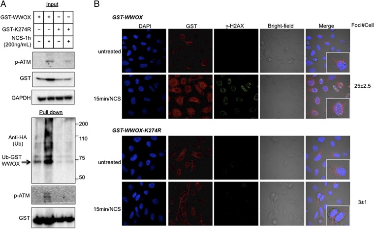
Fig. 6.
Ubiquitination of WWOX at Lys274 after DNA damage. (A) HEK293 cells were transfected with HA-Ub and GST-WWOX or GST-WWOX-K274R plasmids. At 24 h, cells were treated with NCS (200 ng/mL) for an additional 1 h. Cell lysates were blotted against p-ATM, GST (WWOX), and GAPDH. Pulled down complexes were blotted with anti-HA (Ub), anti-GST (WWOX), and anti–p-ATM antibodies. (B) HeLa cells were transfected with HA-Ub and GST-WWOX or GST-WWOX-K274R plasmids. At 24 h, cells were treated with NCS (200 ng/mL) for 15 min. Immunostaining was performed using anti-GST (red) and anti–p-H2AX (green). GST is also observed in control untreated cells, likely because anti-GST stains endogenous GST. DAPI was used as a marker for nuclei. Cells were examined by confocal microscopy under 60× magnification. Quantification of γ-H2AX foci is shown on the right side as the average foci number per cell ± SEM (P < 0.001).

