Fig. 1.
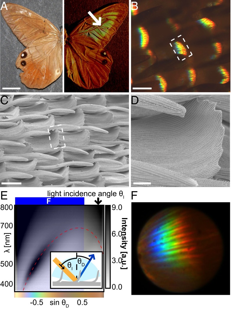
Optical properties of the curled scales in butterfly P. luna. (A) Optical image of P. luna under diffuse lighting (Left) and directional lighting at grazing incidence (Right). Scale bar, 10 mm. (B) Optical micrograph of P. luna scales under oblique illumination. Scale bar, 50 µm. (C) SEM of scales in the colored wing region. Scale bar, 50 µm. White dashed boxes in B and C mark the curled tops of the scales from which the color originates. (D) Close-up image of the curled region of the scale from which the color originates. Scale bar, 20 µm (E) Gray-scale encoded reflection intensity as a function of wavelength and propagation direction showing the inverse color-order diffraction pattern for 65° light incidence. The red dashed line indicates the predicted location of the diffraction due to the cross-rib structures for an orientation of the curled scale sections of −25° relative to the surface normal and a cross-rib periodicity of 390 nm. The blue shaded region signifies the angle range for which the diffraction microscopy image of curled P. luna scales in F was obtained. The color bar under the graph shows the human-eye-perceived color for the spectra observed at the corresponding angles calculated by the CIE 1931 standards (14). (F) Diffraction microscopy image of the colored spot of a P. luna wing showing multiple diffracted orders in similar angular locations due to the variation in the position and angle of the diffracting scales.

