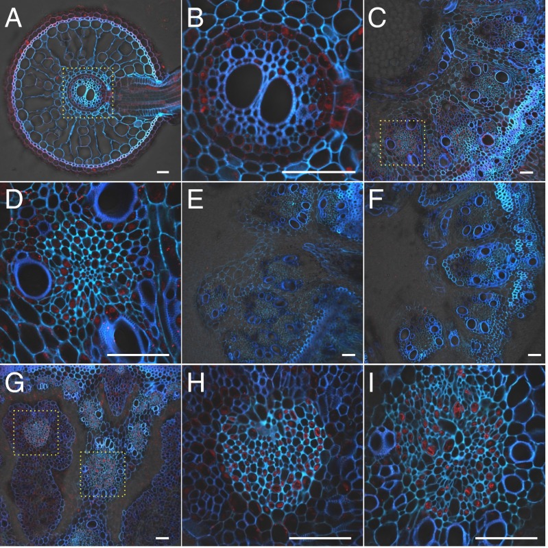
Fig. 3.
Cellular localization of OsABCC1. Immunostaining with an antibody against OsABCC1 was performed in different organs of rice at the vegetative (A–F) and reproductive growth (G–I) stage. (A and B) WT root. (C and D) WT basal node. (E and F) abcc1-1 and abcc1-2 mutant basal node, respectively. (G–I) WT node I. Red color indicates the OsABCC1 antibody-specific signal. Blue color indicates cell wall autofluorescence. Yellow dotted boxes in A, C, and G represent regions magnified in B (root stele), D, H (phloem of EVB), and I (DVB), respectively. (Scale bar, 50 µm.)

