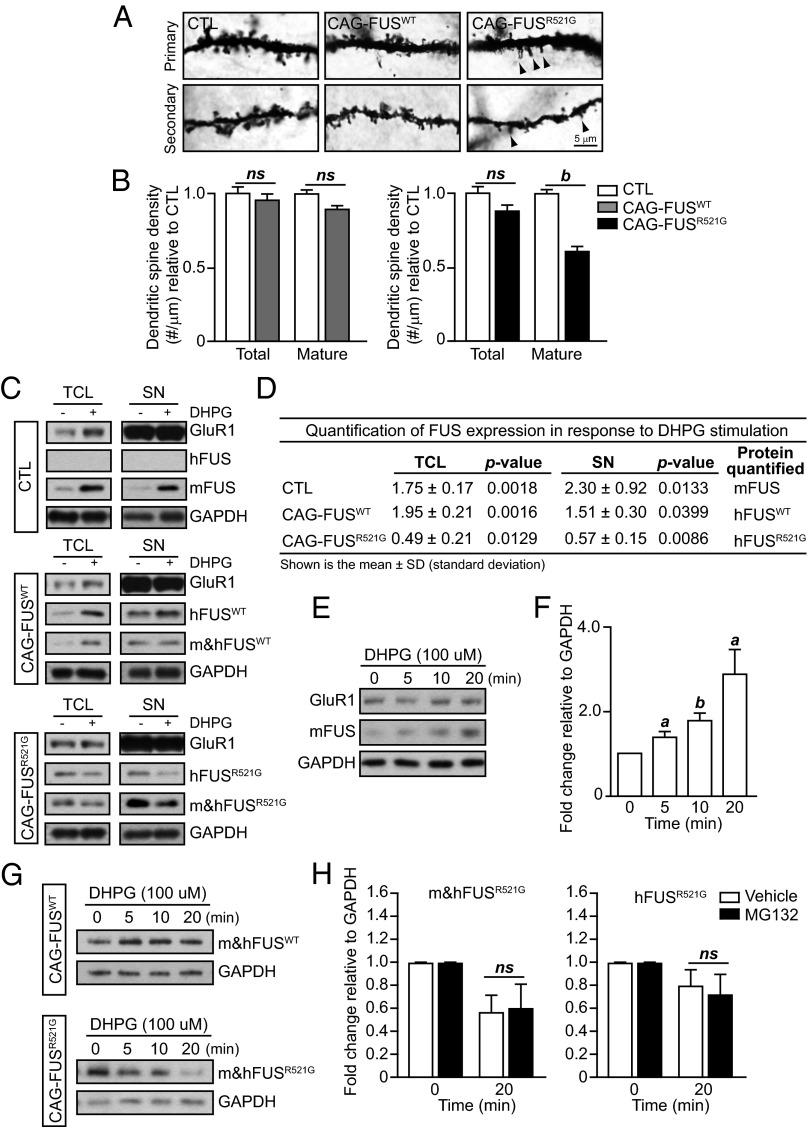
Fig. 5.
Activity-dependent reduction of FUSR521G in response to mGluR stimulation. (A and B) Golgi images of dendritic spines in the apical and basal dendrites of control (CTL), CAG-FUSWT, and CAG-FUSR521G mice. Spine density was analyzed in a total of 30 neurons from cortical layers IV–V in P18 CAG-FUSWT and CAG-FUSR521G mice and their corresponding littermate controls (n = 3 mice per group). In CAG-FUSWT mice the density of mature dendritic spines does not differ in apical and basal dendrites. However, in CAG-FUSR521G mice the density of mature dendritic spines is reduced in the apical and secondary dendrites compared with littermate controls. (We have defined “mature” spines as being mushroom-shaped.) Arrowheads indicate immature spines. (C) Acute cortical tissue slices from littermate P18CTL, CAG-FUSWT, or CAG-FUSR521G mice (n = 3 mice per group) were pretreated with AMPA (20 μM 6,7-dinitroquinoxaline-2,3-dione, DNQX) and NMDA [5 μM 3-(2-carboxypiperazin-4-yl)propyl-1-phosphonic acid, CPP] inhibitors, followed by treatment with 100 μM DHPG for 10 min or no DHPG treatment. Total cell lysates (TCL) and synaptoneurosomes (SNs) were immunoblotted for human FUS (hFUS), mouse FUS (mFUS), total FUS (m&hFUS), GAPDH, and GluR1. GluR1 is enriched in the synaptoneurosome fraction. (D) Quantification of FUS expression from total cell lysates and synaptoneurosomes from acute cortical tissue slices treated with DHPG relative to untreated groups. Immunoblots are representative of three or four separate experiments. P values were obtained by Student t test. (E) Isolated synaptoneurosomes from CTL mice were treated with 100 μM DHPG for the indicated time and were immunoblotted for mFUS, GAPDH, and GluR1. (F) Quantification of FUS relative to GAPDH indicates a significant increase in expression in response to DHPG treatment. (G) Isolated synaptoneurosomes from CAG-FUSWT and CAG-FUSR521G mice were treated with 100 μM DHPG for the indicated time and were immunoblotted for total m&hFUS and GAPDH. Immunoblots are representative of two separate experiments. (H) Isolated synaptoneurosomes from CAG-FUSR521G mice were pretreated with 25 μM MG132 or vehicle (DMSO) before stimulation with 100 μM DHPG for 20 min. Synaptoneurosome lysates were immunoblotted, and m&hFUS and hFUS levels were quantified relative to GAPDH. MG132 did not inhibit the decrease in FUS expression. (B, D, F, and H) ns, not significant. a, P < 0.05; b, P < 0.01 (Student t test). The FUS Santa Cruz antibody was used for all blots for m&hFUS. Three animals were used in each experimental group. (B and H) Error bars represent SEM; (F) Error bars represent SD.

