
Fig. 1.
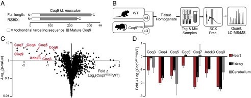
Coq9R239X mice have decreased steady-state levels of CoQ biosynthesis proteins. (A) Coq9R239X truncation scheme designed by García-Corzo et al. (11). (B) Experimental design for proteomic LC-MS/MS analysis of WT and Coq9R239X mice tissue (heart, kidney, cerebellum with n = 3 for each tissue). (C) Volcano plot of protein abundance (•) changes between WT and Coq9R239X mouse heart [Fold Δ Log2(Coq9R239X/WT) versus –Log10(P value)] with COQ proteins labeled. (D) COQ protein abundance changes between WT and Coq9R239X in ( ) heart, (■) kidney, and (
 ) cerebellum [Fold Δ Log2(Coq9R239X/WT)] with error bars showing standard deviation. See also Fig. S1.