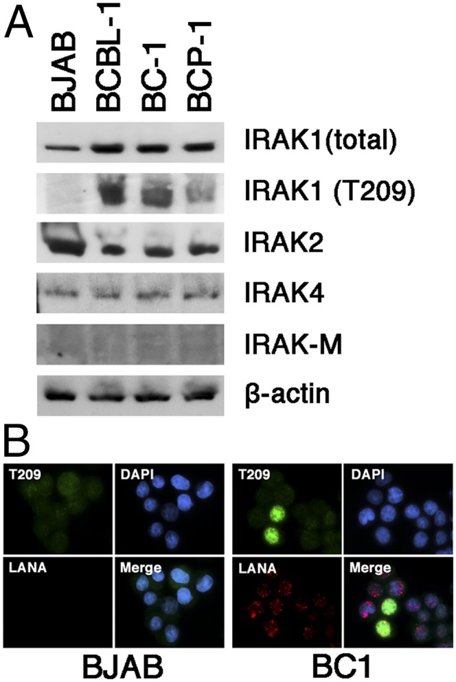
Fig. 4.

Expression of IRAK isoforms in PEL. (A) Shown is a Western blot of multiple PEL samples (BCBL-1, BC-1, BCP-1) and the BJAB Burkitt lymphoma cell line for total IRAK1, phosphoIRAK1T209, IRAK2, IRAK4, IRAK-M, and beta actin. (B) Immunofluorescence of IRAK1 phosphorylation in BJAB and BC1 cells, using antibodies against the T209 phosphorylation site in IRAK1 (green) and LANA (red). Nuclear DNA is counterstained with DAPI (blue).
